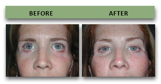
Cranio-orbital Deformities Before and After Photos
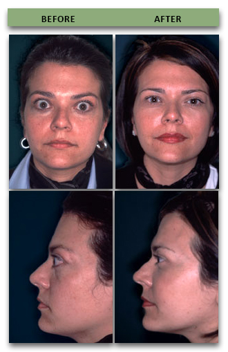
This Graves Disease patient (overactive thyroid gland) suffered from severely swollen eyes. She was unable to close her eyes and lived in constant pain. By increasing the volume of her eye sockets and building up her orbital rims, Dr. Yaremchuk both improved her appearance and eliminated her eye pain. Her surgery was featured in the Health and Science Section of the April 30, 2002 edition of the Boston Globe.
Learn more about this Grave's Disease Patient here.
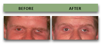
This patient suffered severe head and eye socket fractures resulting in sinking in of his left eye (enophthalmos). Dr. Yaremchuk reconstructed the patient's eye socket roof, walls and floor in order to reposition the eye.
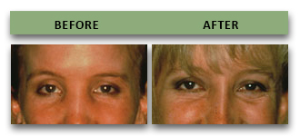
This patient suffered from severe eye socket (nasoethmoidal) fractures, which increased the distance between her eyes, destroyed the bridge of her nose and caused her right eye to sink in (enophthalmos). Dr. Yaremchuk corrected these problems by moving the eye socket walls, reconstructing the nose bridge and adjusting the internal socket walls.
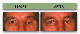
This patient suffered from a severe facial fracture (hemi-nasoethmoidal), which resulted in a misaligned eyed and the sinking of his left eyeball (enophthalmos). Dr. Yaremchuk repositioned and reconstructed the eye socket walls to correct the patient's appearance.
This woman had several attempts to correct the post-traumatic deformity which included bilateral ectropion. Dr. Yaremchuk augmented the temporal deficiency. Osteotomized and moved the zygomatic arch and corrected the ectropion with midface elevation and lateral canthopexy.